Welcome to our Vagus Nerve Anatomy guide!

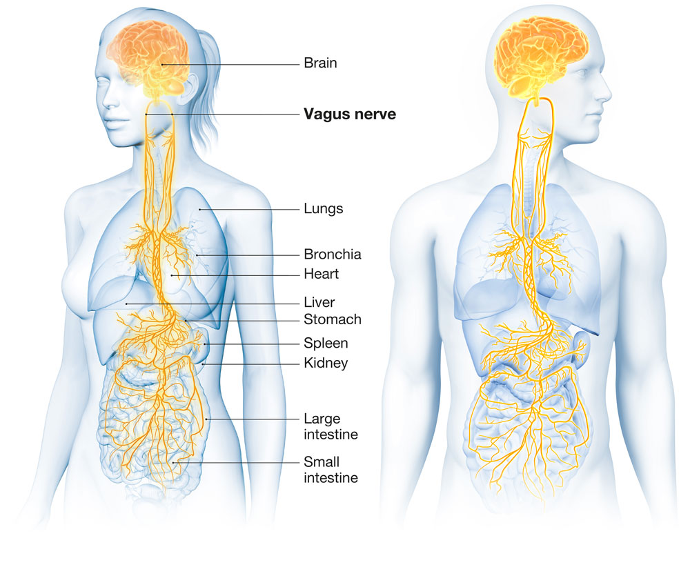
This overview will provide insights into the route taken by the vagus nerve as it traverses the human body.
Join us as we follow the intricate pathways of this wonderous nerve. The medical images provided on this platform or within this content are intended for informational and educational purposes only. They are not a substitute for professional medical advice, diagnosis, or treatment.

Anatomical Structure of the Vagus Nerve

The Vagus Nerve, known as the tenth cranial nerve, is a remarkable anatomical structure characterized by its lengthy composition. It comprises both afferent (sensory, accounting for 80% of its fibers) and efferent (motor, making up 20%) fibers, giving it the appearance of a delicate, whitish vine or a flexible fiber optic cable. This intricate nerve network exhibits a somewhat lacy or feathery appearance, much like the intricate roots of a spreading tree, owing to its numerous branches and smaller nerve fibers. Afferent neurons, a crucial component of the vagus nerve, transmit sensory information from stimuli to the central nervous system and the brain, while efferent neurons carry neural signals away from the central nervous system to muscles, enabling movement and bodily function regulation.

  • 1. Origin: Whilst exiting the lower part of the brainstem, the vagus nerve originates in the medulla oblongata.
  • 2. Ganglia: This ganglion is located in the jugular foramen, which is a bony canal at the base of the brainstem where the vagus nerve exits the cranial cavity. There are two ganglia on the vagus nerve, namely the superior and inferior ganglia.
  • 3. Joining Nerve: The spinal accessory nerve (CN XI) joins the vagus nerve to the inferior ganglion, situated away from the centre of the body at point of attachment.
  • 4. Carotid Sheath: The vagus nerve moves down from the head inside a special tube called the carotid sheath. This tube is found behind and to the side of the internal and common carotid arteries and situated middle to the internal jugular vein.
  • 5. Right Vagus Nerve: The right vagus ganglia travels down and forward near the subclavian artery, then goes behind the innominate artery and downwards to the chest (thoracic cavity). Finally, it joins with the left vagus nerve at the esophageal plexus.
  • 6. Left Vagus Nerve: The left vagus nerve goes forward near the subclavian artery, goes into the chest (thoracic cavity) between the left common carotid and subclavian arteries and then moves down behind the phrenic nerve. Finally, it joins with the right vagus nerve at the esophageal plexus.
  • 7. Neck Branches: Within the neck, the vagus nerve has four branches: pharyngeal branches, superior laryngeal nerve, recurrent laryngeal nerve and superior cardiac nerve.
  • 8. Pharyngeal Branches: These are smaller nerve branches that attach to the muscles in the pharynx (the back of the throat) and the palate (the roof of the mouth).
  • 9. Superior Laryngeal Nerve: It divides into two branches near the bone in your neck called the hyoid bone. The internal branch attaches to your voice box (larynx) and the external branch travel alongside your thyroid blood vessels in your neck.
  • 10. Recurrent Laryngeal Nerves: The right recurrent laryngeal nerve branches near the right subclavian artery and enters the larynx between the cricopharyngeal muscle and the esophagus, while the left recurrent laryngeal nerve loops around the aortic arch to reach the larynx.
  • 11. Cardiac Nerve: While located within the carotid sheath, the vagus nerve releases the superior cardiac nerve, which carries parasympathetic fibres and travels to the heart.
  • 12. Bronchial Branches: The vagus nerve sends out two sets of bronchial branches to the lungs. The front bronchial branches situated front of the lungs form the anterior pulmonary plexus and the back bronchial branch situated back of the lungs form the posterior pulmonary plexus.
  • 13. Esoghageal Plexus: Connected to the fibrous wall of the esophagus, the vagus nerve branches into a web-like structure that envelopes the esophagus. As it descends, the esophageal plexus reaches the lower esophageal sphincter (LES), the opening to the stomach.
  • 14. Gastric Branches: These branches extend from the vagal trunk and create a network of smaller branches, forming a mesh-like structure that envelopes the back and most of the stomach.
  • 15. Celiac Branches: Mainly derived from the right vagus nerve, celiac branches connect to the pancreas, spleen, kidneys, adrenals and small intestine.
  • 16. Hepatic Branches: As it travels deeper into the abdomen, the vagal trunk moves toward the liver and gallbladder, where it splits into smaller branches known as the Hepatic Branches.
  • 17. Renal Branches: While the renal plexus attached to the kidney’s primarily responds and is made up of mostly sympathetic signals, it can still indirectly impact kidney function through its parasympathetic fibres.
  • 18. Pelvic Region: The splanchnic nerve, extending from the vagal trunk, connects to the bladder wall and detrusor muscle, while the pudendal nerve, also originating from the vagal trunk, extends to the genitals, anal sphincters and perineum.

Did you know your vagus has its own signalling neurotransmitter and it's called Acetylcholine (ACh)?

The vagus nerve depends solely on ACh for communication with its assigned organs. This neurotransmitter is like the ultimate messenger in your central nervous system (CNS), ensuring signals get from point A to point B. Interestingly, it has different roles depending on which side of the CNS it's traveling on – whether it's the sympathetic or parasympathetic side, where the vagus nerve resides.

Now, let's discuss the production of ACh. It doesn't magically appear; rather, it undergoes a fascinating metabolic process. Acetylcholine is a combination of acetyl coenzyme A (acetyl-CoA) from your body's energy factories and importantly, choline from your diet. Choline takes a journey from your stomach through your bloodstream, with its destination being the nerve cells of the Vagus Nerve. Within the nerve cell, choline combines with Acetyl-CoA, facilitated by the enzyme Choline Acetyltransferase (ChAT), to create the neurotransmitter Acetylcholine.

So how do we get more of this amazing neurotransmitter. Boosting acetylcholine naturally involves a nutrient-rich diet with choline-rich foods, B-vitamins and omega-3 fatty acids. Antioxidant-rich foods, regular physical exercise, adequate sleep and stress management also contribute to a healthy supply of ACh. Consider acetylcholine precursors like choline supplements and explore herbal support with bacopa monnieri and huperzine A. Limit alcohol and caffeine intake and engage in brain-stimulating activities for overall cognitive health. Always consult a healthcare professional before adding supplements.

Another intriguing fact about Acetylcholine is that once it is created it doesn't just hang around idly. It's stored within nerve cells, ready to make its grand entrance to send signals to target organs. But here's the twist – after it's done its job, ACh doesn't stick around forever. Enter acetylcholinesterase (AChE), the cleanup crew. AChE breaks down Ach back into acetyl-CoA and choline, which isn't discarded but recycled. This recycled choline ensures a continuous supply of ACh, keeping the neural message flow smooth and uninterrupted.

So, the next time you're marvelling at the wonders of your smartphone, remember this: your brain and your vagus nerve have their very own direct messaging system called Acetylcholine, ensuring a seamless flow of signals through your vagus nerve to its target organs.


The Following are images that have been collated from The Netter Collection - one of the most comprehensive collection of hand-painted medical illustrations available with a 50+ year history and a brand identity recognised and accepted as the standard by anyone who has ever received some form of healthcare training.

Illustration of Vagus Nerve (X)

Netter Images. Schema from the Netter Collection.

Learn more...
Illustration of Vagus Nerve (X)

Netter Images. Schema from the Netter Collection.

Learn more...
Cervical Plexus

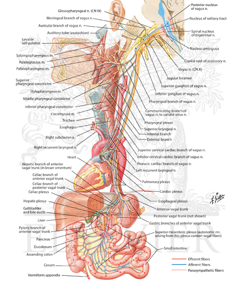
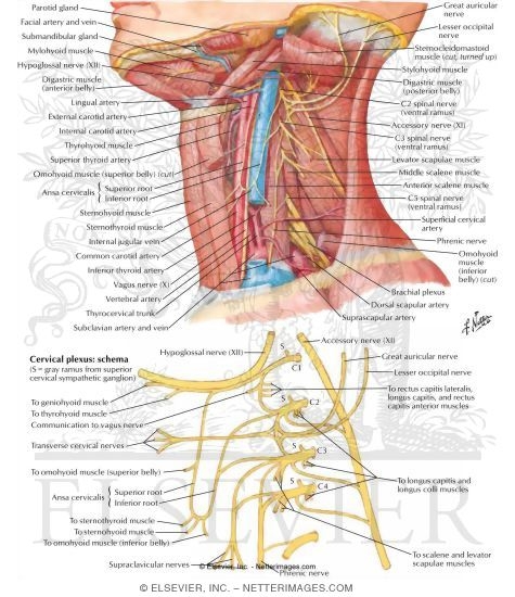
Illustration of Cervical Plexus In Situ Nerves of the Upper Extremity: Cervical Plexus from the Netter Collection

Learn more...
Laryngeal Nerve    

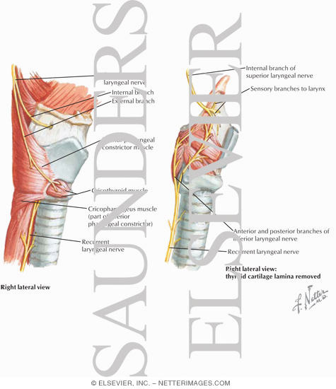
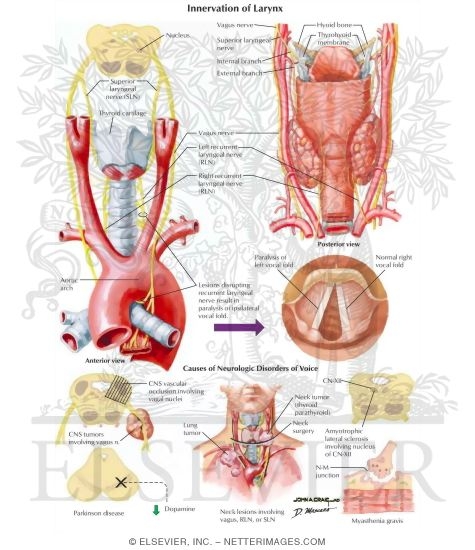
Illustration of Motor and Sensory Branches From the Vagus Nerve from the Netter Collection

Learn more...
The Larynx    

Illustration of Innervation of Larynx from the Netter Collection

Learn more...
The Heart

Illustration of Nerves of Heart from the Netter Collection

Learn more...
The Stomach and Duodenum

The Stomach and Duodenum from the Netter Collection

Learn more...
The Stomach and Duodenum

The Stomach and Duodenum from the Netter Collection

Learn more...
Abdomen and of Perineum

The Stomach and Duodenum from the Netter Collection

Learn more...
Kidneys, Ureters and Bladder

From the Netter Collection

Learn more...
Disclaimer: The information provided on this webpage is intended for general informational purposes only and should not be considered a substitute for professional medical advice, diagnosis, or treatment. Always seek the advice of your physician or other qualified healthcare provider with any questions you may have regarding a medical condition. Read full Disclaimer...